Clinical workflow
A lens wearer presents with a painful red eye.
OptoGuide™ gives the contact lens red eye its own safety pathway — capture the corneal danger cues, separate infective from sterile causes, and draft a same-day referral when keratitis is a concern.
Structured for Australian optometry practice. Source-cited clinical reference; not individually clinician-reviewed.
Clinical decision support only
Red flags: assess urgently before anything else
Stop lens wear immediately. Same-day corneal review is supported when infection risk and corneal signs are both present.
- Moderate pain or marked photophobia.
- Focal staining, an infiltrate, or corneal opacity.
- Reduced acuity or central corneal involvement.
- Overnight wear or water exposure before symptoms.
- A unilateral acute presentation that does not look superficial.
One connected workflow, not separate lookups
Recognition, management, prescribing, and referral usually live in different tools. In OptoGuide™ they are one path, and each step hands off to the next so the decision keeps moving.
Step 1
Enter the contact lens pathway
Start from a dedicated contact-lens red-eye front door rather than a generic red-eye checklist, so lens history and corneal risk are captured from the outset.
- Stop lens wear immediately while you assess the cause.
- Record wear pattern, overnight wear, and any water exposure.
- Measure acuity and localise any corneal involvement.
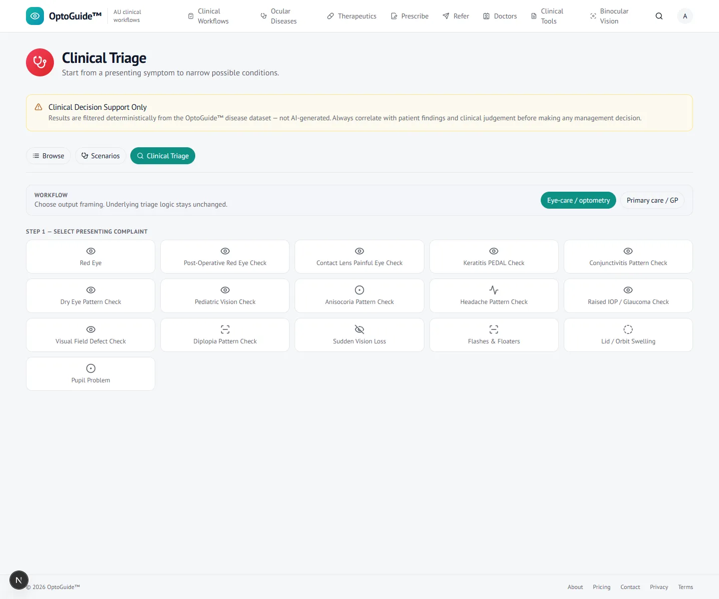
The consult starts from the presenting complaint, not a diagnosis. Step 2
Capture the corneal danger clues
The findings screen captures the keratitis-risk cues — epithelial defect, infiltrate, central lesion, anterior chamber reaction — while keeping patient identifiers out of the app.
- Central staining or an infiltrate raises concern for microbial keratitis.
- Separate infective risk from sterile or mechanical causes early.
- A low threshold for referral applies in high-risk wearers.
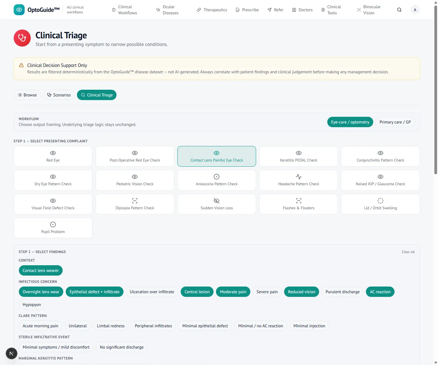
Step 3
Keep keratitis visible
A deterministic result keeps microbial keratitis in view instead of buried in a red-eye list, and distinguishes it from sterile infiltrative or intolerance patterns.
- Infectious concern is surfaced early and clearly.
- The reasoning is transparent so you can correlate it with the cornea.
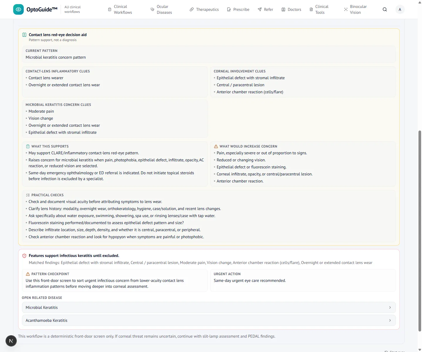
Step 4
Open the keratitis workflow
Open the microbial keratitis workflow for structured recognition, steroid caution, escalation, and follow-up-failure logic.
- Escalate rather than observe when the diagnosis is uncertain in a high-risk wearer.
- Document corneal signs, lens history, and acuity before escalation.
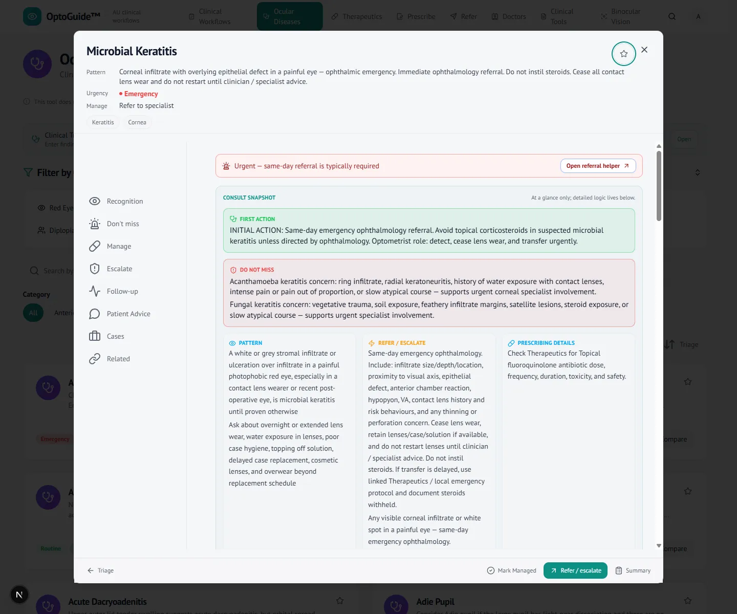
Step 5
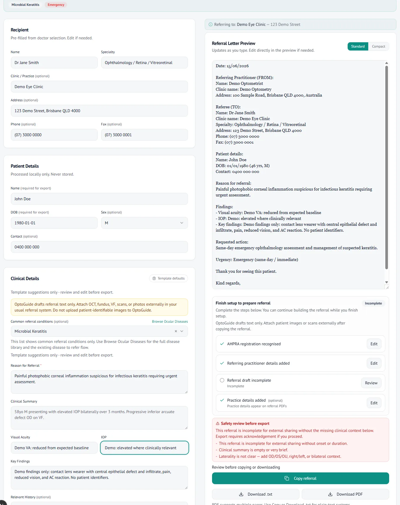
Draft a same-day corneal referral
Draft the referral from the disease context with urgency prefilled, while the findings stay clinician-entered. The letter is generated for you to review and copy.
- Same-day corneal / ophthalmology wording for suspected microbial keratitis.
- Copy-first output for Oculo, Medical Objects, or your PMS. No patient data is stored.
Clinical basis
This guidance reflects standard optometric clinical reasoning based on:
- Australian optometry clinical practice patterns
- Australian medicines regulation and PBS prescribing context
- Common ophthalmology referral standards
- Evidence-based clinical training and practice
Give the next contact lens red eye a safety-first pathway.
Free 14-day full-access trial. No credit card required.