Clinical workflow
A lens wearer with a corneal infiltrate.
The question is not just “keratitis or not” — it is whether this is a sterile contact-lens peripheral ulcer or a microbial keratitis. OptoGuide™ keeps the discriminating features side by side and supports escalation rather than observation when the picture is uncertain.
Structured for Australian optometry practice. Source-cited clinical reference; not individually clinician-reviewed.
Clinical decision support only
Red flags: assess urgently before anything else
Stop lens wear immediately, whichever way the diagnosis falls. These features move the picture away from a sterile peripheral ulcer toward microbial keratitis and support same-day emergency ophthalmology.
- A central or paracentral infiltrate, or one that is large rather than small and well-defined.
- Dense staining or a significant epithelial defect over the infiltrate.
- Severe pain out of proportion to the lesion, marked photophobia, or reduced vision.
- A significant anterior chamber reaction or hypopyon.
- An infiltrate that is worsening or failing to improve, or a ring infiltrate forming — with water exposure in lenses, raises concern for Acanthamoeba.
- Overnight wear, or a delay in removing lenses despite symptoms.
One connected workflow, not separate lookups
Recognition, management, prescribing, and referral usually live in different tools. In OptoGuide™ they are one path, and each step hands off to the next so the decision keeps moving.
Step 1
Read the infiltrate pattern
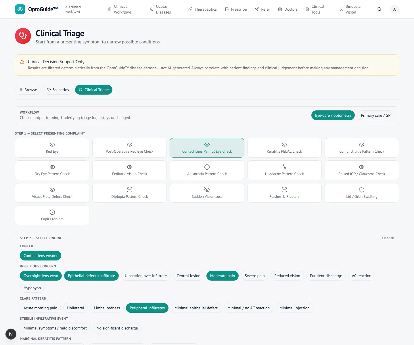
Bring the exposure history and the corneal signs together from the outset. The size, position, staining, and chamber reaction are what separate a sterile peripheral ulcer from an infective one.
- Stop lens wear immediately — this is mandatory whichever way the diagnosis falls.
- A sterile peripheral ulcer is typically small and well-defined, sits peripherally with a clear limbal zone, and stains minimally.
- Pain that is mild relative to the size of the infiltrate supports a sterile picture; pain out of proportion does not.
- Record overnight wear, water exposure, and any delay in removing lenses.
Step 2
Open the matching condition
Open the condition for structured recognition, antibiotic-cover caution, close-review expectations, and escalation logic. Use a low threshold for referral in high-risk wearers.
- Escalate rather than observe if the diagnosis is uncertain in a high-risk wearer.
- Chloramphenicol is not appropriate first-line cover for a contact-lens-related infiltrate — Pseudomonas risk means Gram-negative cover is needed. Check Therapeutics for the current choice and prescribing detail.
- Review closely to confirm infiltrate size, epithelial status, vision, and anterior chamber stay stable or improving.
- Do not resume lens wear until the infiltrate has fully resolved.

Step 3
Escalate with a same-day referral
When infection risk and corneal signs are both present, draft a same-day corneal referral in the same flow. The letter is generated for you to review and copy — OptoGuide never sends it.
- Escalate if the lesion is worsening or not improving, central or paracentral, larger, densely staining, or accompanied by marked pain, reduced vision, or chamber activity.
- A non-resolving infiltrate with a ring pattern or a history of water exposure raises concern for Acanthamoeba — refer as an emergency.
- Same-day corneal / ophthalmology wording for suspected microbial keratitis.
- For recurrent episodes, address lid carriage, move to daily disposable lenses, and stop overnight wear.
- Copy-first output for your existing referral systems. No patient data is stored.
Clinical basis
This guidance reflects standard optometric clinical reasoning based on:
- Australian optometry clinical practice patterns
- Australian medicines regulation and PBS prescribing context
- Common ophthalmology referral standards
- Evidence-based clinical training and practice
Give the next high-risk lens wearer a low threshold to escalate.
Free 14-day full-access trial. No credit card required.