Management workflow
Blepharitis and MGD: foundations beat drops.
OptoGuide™ structures triage and management so the plan is in front of you — lid hygiene and warm compresses as the foundation, with clear cues for when to escalate.
Structured for Australian optometry practice. Source-cited clinical reference; not individually clinician-reviewed.
Clinical decision support only
Red flags: assess urgently before anything else
Uncomplicated blepharitis is bilateral, chronic, and optometrist-managed. Unilateral, refractory, or destructive lid-margin change supports referral for eyelid malignancy assessment rather than repeated hygiene courses.
- A corneal infiltrate, epithelial defect, significant pain, photophobia, or reduced vision — raises concern for microbial keratitis.
- A peripheral corneal infiltrate alongside chronic lid-margin disease — raises concern for marginal keratitis.
- Unilateral, persistent, or refractory lid-margin disease rather than a bilateral chronic pattern.
- Lash loss, lid-margin distortion, ulceration, or a chalazion recurring at the same site.
- Marked focal lid tenderness, spreading swelling, or any concern for cellulitis.
One connected workflow, not separate lookups
Recognition, management, prescribing, and referral usually live in different tools. In OptoGuide™ they are one path, and each step hands off to the next so the decision keeps moving.
Step 1
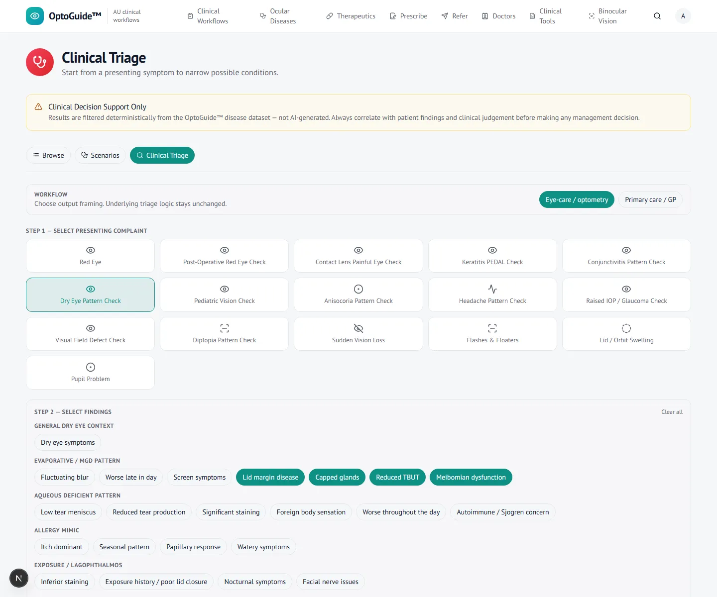
Triage the presentation and findings
Start from the presenting complaint, then the findings — lid-margin telangiectasia, capped or plugged meibomian glands, and a foamy tear film, with grittiness, burning, and intermittent blur.
- Bilateral chronic irritation with lash debris or collarettes points to lid-margin disease rather than acute conjunctivitis.
- Collarettes or cylindrical dandruff at the lash base should trigger a Demodex check.
- Capped glands, turbid meibum, or recurrent chalazia indicate posterior blepharitis / MGD overlap.
- Document lid-margin findings and corneal staining before labelling the case routine.
Step 2
Open the management workflow
Open the condition for structured, optometrist-led management — warm compresses to soften meibum, lid hygiene and lid-margin cleaning, and lubricants for surface instability.
- Foundation therapy — warm compress, lid massage where glands are obstructed, and consistent lid-margin cleansing — is the first-line.
- Explain chronicity: treatment controls flares and recurrence rather than producing a permanent cure after one short course.
- Use a Demodex-directed pathway where collarettes are present, rather than generic hygiene alone.
- Reassess for rosacea, seborrhoeic dermatitis, or contact dermatitis when disease keeps recurring.

Step 3
Know when to escalate
The workflow keeps the escalation cues visible so routine lid-margin disease still has a defined path out when the picture changes. The clinician confirms findings and stays responsible.
- Escalate rather than treating as routine blepharitis if there is a corneal infiltrate, significant staining, severe pain, photophobia, or reduced vision.
- A peripheral infiltrate with chronic lid disease moves the case toward marginal keratitis assessment.
- Reconsider the diagnosis — including eyelid malignancy — when disease is unilateral, refractory, or destroys normal lid architecture.
- Escalate for marked focal lid tenderness, spreading swelling, or a cellulitis concern.
Clinical basis
This guidance reflects standard optometric clinical reasoning based on:
- Australian optometry clinical practice patterns
- Australian medicines regulation and PBS prescribing context
- Common ophthalmology referral standards
- Evidence-based clinical training and practice
Put the lid-disease plan in front of you at the point of care.
Free 14-day full-access trial. No credit card required.