Clinical workflow
A patient notices one pupil looks bigger.
OptoGuide™ gives pupil problems a structured neuro-ophthalmic front door — use lighting pattern and red flags to separate benign anisocoria from the dangerous pupils that need urgent assessment.
Structured for Australian optometry practice. Source-cited clinical reference; not individually clinician-reviewed.
Clinical decision support only
Red flags: assess urgently before anything else
Any CN III pattern with even partial pupil involvement supports emergency assessment for compressive or aneurysmal pathology — hours matter. Do not assume a microvascular cause because the patient is diabetic.
- A dilated pupil that reacts poorly, with ptosis and a “down-and-out” eye — limited adduction, elevation, and depression.
- Even partial pupil involvement — examine in dim light, because slight anisocoria is still significant.
- Acute onset with severe headache, periorbital or retro-orbital pain, or neck pain.
- New diplopia alongside the pupil difference.
- A newly small pupil with ptosis (a Horner pattern), or a history of trauma or possible pharmacologic exposure.
One connected workflow, not separate lookups
Recognition, management, prescribing, and referral usually live in different tools. In OptoGuide™ they are one path, and each step hands off to the next so the decision keeps moving.
Step 1
Start with the pupil problem
Enter from the presenting complaint. The pathway prompts the observations that actually change urgency — onset, lighting behaviour, and associated lid or motility signs.
- Note whether the difference is greater in bright or dim light.
- Look for ptosis, a motility limitation, or diplopia alongside the pupil.
- Establish onset — sudden change is treated differently from a long-standing difference.
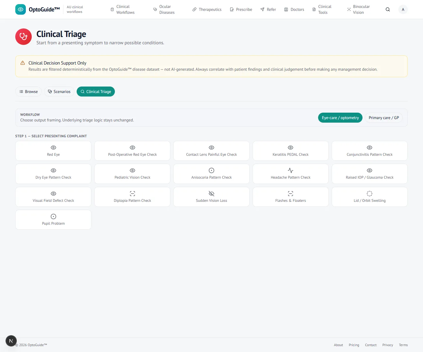
The consult starts from the presenting complaint, not a diagnosis. Step 2
Use lighting pattern plus red flags
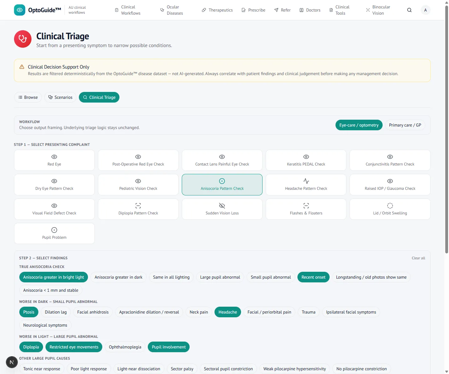
Capture the lighting-pattern and safety cues together. The screen keeps the dangerous combinations — acute onset, ptosis, motility loss, headache — in view rather than defaulting to a benign explanation.
- Greater in bright light means the larger pupil is the abnormal one — the pathway that leads toward CN III.
- Greater in dim light means the smaller pupil is the abnormal one — the pathway that leads toward Horner syndrome.
- Acute onset with ptosis and a motility deficit is treated as a red-flag combination.
- Check the lids and motility in the same breath as the pupil — the combination is what changes urgency.
Step 3
Separate benign from dangerous
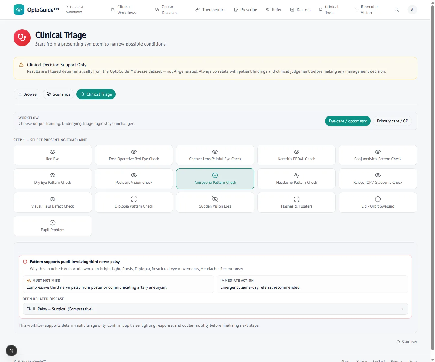
A rule-based result keeps benign physiologic anisocoria distinguishable while dangerous, pupil-involving patterns are escalated — no probabilistic guessing.
- A physiologic pattern is small, long-standing and stable, similar in bright and dim light, with normal pupil reactions, iris appearance and motility — old photographs often settle it.
- New or changing anisocoria, or any ptosis, diplopia, motility deficit, headache, neck pain or iris abnormality, is not a physiologic pattern.
- Pupil-involving third nerve and Horner patterns are held visible for escalation.
- Pharmacologic and traumatic causes stay on the list rather than being assumed away.
Step 4
Open the danger workflow
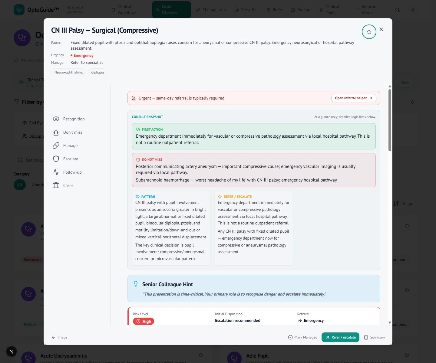
Open the fitting condition for structured recognition and escalation framing. A pupil-involving third nerve palsy is treated as time-critical.
- Recognition and escalation guidance are presented conservatively — no diagnosis is asserted.
- The workflow points to the urgency of assessment, not a specific cause.
Step 5
Escalate with a clear referral
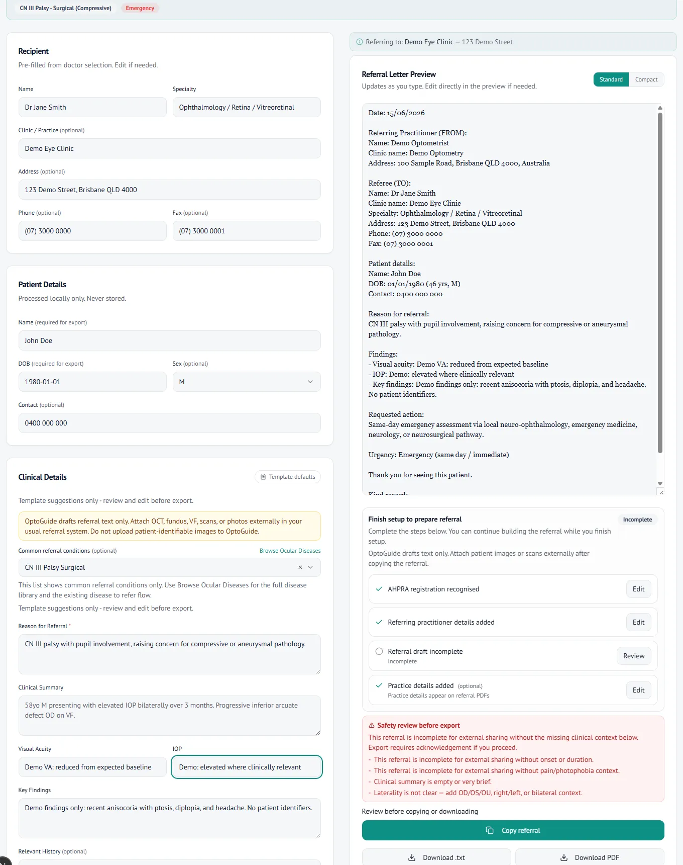
When the picture is neurologically suspicious, draft an urgent referral in the same flow. The letter text is generated for you to review and copy — OptoGuide never sends it.
- Urgency wording is matched to the suspected pattern.
- Copy-first output for your existing referral systems. No patient data is stored.
Clinical basis
This guidance reflects standard optometric clinical reasoning based on:
- Australian optometry clinical practice patterns
- Australian medicines regulation and PBS prescribing context
- Common ophthalmology referral standards
- Evidence-based clinical training and practice
Give the next unequal pupil a structured front door.
Free 14-day full-access trial. No credit card required.